
Types of Mycoplasma testing
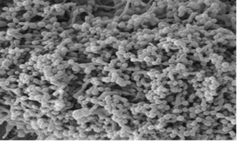
Mycoplasma covering the surface of a single Vero cell in culture
Mycoplasma are the smallest free-living microorganisms, they cannot be seen using phase contrast microscopy or enriched in the standard sterility test method detailed above. They are common contaminants of cell cultures (Mycoplasma contamination of cell cultures: Incidence, sources, effects, detection, elimination, prevention - PubMed.) and effects of mycoplasma contamination on cell lines can include:
-
alteration of growth rate
-
induction of morphological changes
-
chromosomal aberrations
-
altered cell metabolism
-
decreased viability upon resuscitation of frozen ampoule.
-
induction of transformation, apoptosis, cytokines, signal transduction and oxidative radicals
-
macrophage activation
-
inhibition of antigen presentation
Sources of Mycoplasma in Cell Culture
-
Mycoplasma infections can originate from donor tissues. For example, M. orale infection can be found in cell lines from the respiratory tract and M. fermentans in cell lines from urino-genital tract
-
Mycoplasma can be acquired in culture. For example, M. arginini, M. hyorhinis, A. laidlawii can originate in serum or culture medium and reagents
-
Other sources include other infected cell lines and laboratory staff (M. orale, M. fermentans)
-
M. hyorhinis has been associated with poor quality trypsin.
-
Other animal reservoir Mycoplasma can persist in the environment (e.g. M. gallisepticum from chickens can persist in water)
Tests Provided by ECACC
It is essential to know which test is needed.
|
Test |
Sensitivity |
Detection Range |
|---|---|---|
|
Hoechst Stain |
Medium |
all species |
|
Culture Isolation |
High |
all except M. hyorhinis strains |
|
qPCR |
High |
52 species including 6 most common cell culture contaminants (Drexler et al., 2002, Cytotechnology 39: 75–90, 2002) |
ISO 17025:7017 accredited tests provided by ECACC QC Team to Detect Mycoplasma in Cell Culture samples
The Guidelines for the use of cell lines in biomedical research - PubMed. gives guidance on how to select the best test method for your research. It is good practice to test with two or more complementary methods, taking into account the sensitivity and detection range of the tests as given in the table above and the risk associated with your sample (e.g. master cell banks will benefit from additional testing as they will be used to make further banks). European Pharmacopeia describes Indirect DNA (Hoechst) Stain, Culture Isolation and Nucleic Acid Amplification (PCR) techniques in chapter 2.6.7. The FDA removed the 21 CFR 610.30 requirement for either Indirect DNA (Hoechst) Stain or Culture Isolation to allow for more flexibility in determining which test to use.
1. qPCR
This assay consists of a closed tube qPCR detection format and uses TaqMan probes and primers derived from the 16SrRNA region; these were designed manually in-house and consist of three sense primers and four TaqMan probes targeted to anneal to specific regions common in many bacteria.
Specificity of the assay is conferred by the 53 anti-sense primers used, which overlap a broadly conserved region in the 16SrRNA gene of mycoplasma but differ significantly from other (non-mycoplasma) bacterial sequences to enable differentiation from phylogenetically related bacteria. The assay also incorporates an inhibition control that is detected in parallel to the mycoplasma target in a duplex, single-tube reaction.
The assay has been validated as detecting less than 10 genomic copies of the following species:
|
No. |
Species |
|---|---|
|
1 |
Acholeplasma laidlawii |
|
2 |
Mycoplasma arginini |
|
3 |
Mycoplasma fermentans |
|
4 |
Mycoplasma gallisepticum |
|
5 |
Mycoplasma hyorhinis |
|
6 |
Mycoplasma orale |
|
7 |
Mycoplasma pneumoniae |
|
8 |
Mycoplasma synoviae |
|
9 |
Acholeplasma axanthum |
|
10 |
Acholeplasma equifetale |
|
11 |
Acholeplasma granularum |
|
12 |
Acholeplasma hippikon |
|
13 |
Acholeplasma modicum |
|
14 |
Acholeplasma morum |
|
15 |
Acholeplasma oculi |
|
16 |
Acholeplasma parvum |
|
17 |
Entomoplasma ellychniae |
|
18 |
Entomoplasma melaleucae |
|
19 |
Mesoplasma entomophilum |
|
20 |
Mesoplasma florum |
|
21 |
Mycoplasma alvi |
|
22 |
Mycoplasma anatis |
|
23 |
Mycoplasma arthritidis |
|
24 |
Mycoplasma bovirhinis |
|
25 |
Mycoplasma bovoculi |
|
26 |
Mycoplasma buccale |
|
27 |
Mycoplasma californicum |
|
28 |
Mycoplasma canadense |
|
29 |
Mycoplasma canis |
|
30 |
Mycoplasma citelli |
|
31 |
Mycoplasma conjunctivae |
|
32 |
Mycoplasma cricetuli |
|
33 |
Mycoplasma dispar |
|
34 |
Mycoplasma edwardii |
|
35 |
Mycoplasma gateae |
|
36 |
Mycoplasma glycophilum |
|
37 |
Mycoplasma hominis |
|
38 |
Mycoplasma hyopharyngis |
|
39 |
Mycoplasma hyopneumoniae |
|
40 |
Mycoplasma lipophilum |
|
41 |
Mycoplasma maculosum |
|
42 |
Mycoplasma muris |
|
43 |
Mycoplasma opalescens |
|
44 |
Mycoplasma ovipneumoniae |
|
45 |
Mycoplasma pulmonis |
|
46 |
Mycoplasma salivarium |
|
47 |
Mycoplasma spumans |
|
48 |
Mycoplasma verecundum |
|
49 |
Ureaplasma canis |
|
50 |
Ureaplasma diversum |
|
51 |
Ureaplasma urealyticum |
|
52 |
Spiroplasma citri |
The assay was also validated as not detecting closely related Lactobacilli, Clostridia and Streptococcal species. This test is rapid and highly sensitive and suitable for in-process testing and final testing of working and master cell banks.
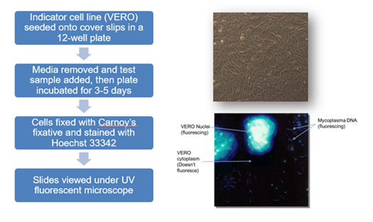
2. Indirect Hoechst Stain
This test is used to detect mycoplasma by Hoechst stain using an indicator cell line (Vero ECACC 84113001) and meets the requirements of the FDA Points to Consider 1993.
The test sample is inoculated onto Vero cells which have been grown on coverslips in tissue culture dishes. Both negative and positive controls (Mycoplasma hyorhinis NCTC 10112 and Mycoplasma orale NCTC 10130) are also set up. All samples are then incubated for 3-5 days after which the samples are fixed to the coverslip and stained using the fluorescent dye (Hoechst 33258). The fixed samples are then examined under UV fluorescence at x100 magnification. Positive samples are identified by their characteristic particulate or filamentous pattern of fluorescence on the cell surface or if contamination is heavy in the surrounding area. Negative samples demonstrate no evidence of fluorescing DNA outside of the cell membrane against the fluorescing Vero cell nuclei.
3. Culture Isolation
This test is used to detect Mycoplasma by Culture Isolation and meets the requirements of the FDA Points to Consider 1993.
This is sometimes referred to as the gold standard method due to its high sensitivity arising from enriching any Mycoplasma present and broad detection range, limited only by the medium used and the inability of some Mycoplasma species to grow on solid medium.
Due to the long incubation times associated with growing Mycoplasma, this test is usually reserved for high-risk samples and Master Cell banks.
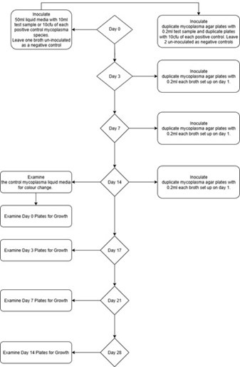
Process Flow for ECACC Culture Isolation Test